Cynomolgus / Rhesus Fractalkine / CX3CL1 Protein (His Tag)
Fractalkine, CX3CL1
- 100ug (NPP4422) Please inquiry
| Catalog Number | P90316-C08H |
|---|---|
| Organism Species | Cynomolgus |
| Host | Human Cells |
| Synonyms | Fractalkine, CX3CL1 |
| Molecular Weight | The recombinant cynomolgus CX3CL1 consists of 87 amino acids and predicts a molecular mass of 10.1 kDa. |
| predicted N | Gln 25 |
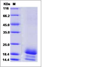
| SDS-PAGE |  |
| Purity | > (71.4+6.5+20.3) % as determined by SDS-PAGE. |
| Protein Construction | A DNA sequence encoding the cynomolgus CX3CL1 (EHH60415.1) (Met1-Gly100) was expressed with a polyhistidine tag at the C-terminus. Cynomolgus and Rhesus CX3CL1 sequences are identical. |
| Bio-activity | |
| Research Area | Cardiovascular |Atherosclerosis |Vascular Inflammation |Leukocyte recruitment |Chemokines in Leukocyte recruitment |
| Formulation | Lyophilized from sterile PBS, pH 7.4. 1. Normally 5 % - 8 % trehalose, mannitol and 0.01% Tween80 are added as protectants before lyophilization. Specific concentrations are included in the hardcopy of COA. |
| Background | Fractalkine or Chemokine (C-X3-C motif) ligand 1 (CX3CL1) is a member of the CX3C chemokine family. Fractalkine / CX3CL1 is a unique chemokine that functions not only as a chemoattractant but also as an adhesion molecule and is expressed on endothelial cells activated by proinflammatory cytokines, such as interferon-gamma and tumor necrosis factor-alpha. Fractalkine/CX3CL1 is expressed in a membrane-bound form on activated endothelial cells and mediates attachment and firm adhesion of T cells, monocytes and NK cells. Fractalkine / CX3CL1 is associated with dendritic cells (DC) in epidermis and lymphoid organs. The fractalkine receptor, CX3CR1, is expressed on cytotoxic effector lymphocytes, including natural killer (NK) cells and cytotoxic T lymphocytes, which contain high levels of intracellular perforin and granzyme B, and on macrophages. Soluble fractalkine causes migration of NK cells, cytotoxic T lymphocytes, and macrophages, whereas the membrane-bound form captures and enhances the subsequent migration of these cells in response to secondary stimulation with other chemokines. |
| Reference |
