
Cynomolgus / Rhesus c-MET / HGFR Protein (Fc Tag)
MET
- 100ug (NPP1096) Please inquiry
| Catalog Number | P90304-C02H |
|---|---|
| Organism Species | Cynomolgus |
| Host | Human Cells |
| Synonyms | MET |
| Molecular Weight | The recombinant cynomolgus/rhesus MET comprises 1146 amino acids and has a calculated molecular mass of 128.3 KDa. The apparent molecular mass of it is approximately 100.2 and 42.5 KDa in SDS-PAGE under reducing conditions. |
| predicted N | Glu 25 |
| SDS-PAGE | 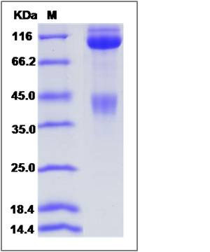 |
| Purity | (66.7+31.5) % as determined by SDS-PAGE |
| Protein Construction | A DNA sequence encoding the cynomolgus/rhesus MET (NP_001162100.1) (Met1-Thr932) was expressed with the Fc region of human IgG1 at the C-terminus. Cynomolgus and Rhesus MET sequences are identical. |
| Bio-activity | Immobilized Cynomolgus HGF (P90286-CNAH) at 10 μg/ml (100 μl/well) can bind Cynomolgus MET-Fc, EC50 of Cynomolgus MET-Fc is 0.04-0.09 μg/ml. |
| Research Area | Cancer |Signal transduction |Protein Phosphorylation |Tyrosine Kinase |Receptor Tyrosine Kinases |
| Formulation | Lyophilized from sterile PBS, pH 7.4. 1. Normally 5 % - 8 % trehalose and mannitol are added as protectants before lyophilization. Specific concentrations are included in the hardcopy of COA. |
| Background | Hepatocyte growth factor receptor (HGFR), also known as c-Met or mesenchymal-epithelial transition factor (MET), is a receptor tyrosine kinase (RTK) that has been shown to be overexpressed and/or mutated in a variety of malignancies. HGFR protein is produced as a single-chain precursor, and HGF is the only known ligand. Normal HGF/HGFR signaling is essential for embryonic development, tissue repair or wound healing, whereas aberrantly active HGFR has been strongly implicated in tumorigenesis, particularly in the development of invasive and metastatic phenotypes. HGFR protein is a multifaceted regulator of growth, motility, and invasion, and is normally expressed by cells of epithelial origin. Preclinical studies suggest that targeting aberrant HGFR signaling could be an attractive therapy in cancer. |
| Reference |