Human PLS3 / Plastin 3 Protein (His Tag)
BMND18,T-plastin
- 100ug (NPP2415) Please inquiry
| Catalog Number | P15042-H07E |
|---|---|
| Organism Species | Human |
| Host | E. coli |
| Synonyms | BMND18,T-plastin |
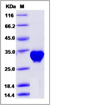
| Molecular Weight | The recombinant human PLS3 consists of 289 amino acids and predicts a molecular mass of 32.2 KDa. It migrates as an approximately 30-34 KDa band in SDS-PAGE under reducing conditions. |
| predicted N | His |
| SDS-PAGE |  |
| Purity | > 95 % as determined by SDS-PAGE |
| Protein Construction | A DNA sequence encoding the human PLS3 (P13797) (Gly102-Asn375) was expressed with a polyhistide tag at the N-terminus. |
| Bio-activity | |
| Research Area | Cardiovascular |Angiogenesis |Adhesion Molecules in Angiogenesis |Extracellular Matrix |Structures |Focal Adhesions | |
| Formulation | Lyophilized from sterile PBS, 10% Glycerol, pH 7.4. 1. Normally 5 % - 8 % trehalose and mannitol are added as protectants before lyophilization. Specific concentrations are included in the hardcopy of COA. |
| Background | PLS3, also known as plastin 3, belongs to the plastin family. Members of this family are actin-binding proteins that are conserved throughout eukaryote evolution and expressed in most tissues of higher eukaryotes. There are two ubiquitous plastin isoforms in humans: L and T. The L isoform is expressed only in hemopoietic cell lineages, while the T isoform has been found in all other normal cells of solid tissues that have replicative potential (fibroblasts, endothelial cells, epithelial cells, melanocytes, etc.). PLS3 contains 2 actin-binding domains, 4 CH (calponin-homology) domains and 2 EF-hand domains. It is expressed in a variety of organs, including muscle, brain, uterus and esophagus. |
| Reference |
