Mouse MAG / GMA / Siglec-4 Protein (ECD, Fc Tag)
Gma,siglec-4a
- 100ug (NPP3415) Please inquiry
| Catalog Number | P51398-M02H |
|---|---|
| Organism Species | Mouse |
| Host | Human Cells |
| Synonyms | Gma,siglec-4a |
| Molecular Weight | The recombinant mouse Mag consists of 735 amino acids and predicts a molecular mass of 81.6 kDa. |
| predicted N | Gly 20 |
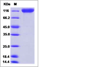
| SDS-PAGE |  |
| Purity | > 95 % as determined by SDS-PAGE. |
| Protein Construction | A DNA sequence encoding the mouse Mag (NP_034888.1) (Met1-Pro516) was expressed with the Fc region of human IgG1 at the C-terminus. |
| Bio-activity | |
| Research Area | Neuroscience |Cell Adhesion Proteins |Membrane Proteins |
| Formulation | Lyophilized from sterile PBS, pH 7.4. 1. Normally 5 % - 8 % trehalose, mannitol and 0.01% Tween80 are added as protectants before lyophilization. Specific concentrations are included in the hardcopy of COA. |
| Background | The myelin-associated glycoprotein (MAG) contains five immunoglobulin-like domains and belongs to the sialic-acid-binding subgroup of the Ig superfamily. MAG is a transmembrane glycoprotein of 100kDa localized in myelin sheaths of periaxonal Schwann cell and oligodendroglial membranes where it functions in glia-axon interactions. It appears to function both as a receptor for an axonal signal that promotes the differentiation, maintenance and survival of oligodendrocytes and as a ligand for an axonal receptor that is needed for the maintence of myelinated axons. MAG contains a carbohydrate epitope shared with other glycoconjugates that is a target antigen in autoimmune peripheral neuropathy associated with IgM gammopathy and has been implicated in a dying back oligodendrogliopathy in multiple sclerosis. MAG is considered as a transmembrane protein of both CNS and PNS myelin and it strongly inhibits neurite outgrowth in both developing cerebellar and adult dosal root ganglion neurons. In contrast, MAG promotes neurite outgrowth from newborn DRG neurons. Thus, MAG may be responsible for the lack of CNS nerve regeneration and may influce both temporally and spatially regeneration in the PNS. |
| Reference |
