Mouse SDPR Protein (aa 2-180, His Tag)
SDPR
- 100ug (NPP3470) Please inquiry
| Catalog Number | P50603-M07E |
|---|---|
| Organism Species | Mouse |
| Host | E. coli |
| Synonyms | SDPR |
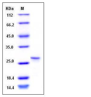
| Molecular Weight | The recombinant mouse SDPR consisting of 186 amino acids and has a calculated molecular mass of 21 kDa. rmSDPR migrates as an approximately 28 kDa band in SDS-PAGE under reducing conditions |
| predicted N | Met |
| SDS-PAGE |  |
| Purity | > 95 % as determined by SDS-PAGE |
| Protein Construction | A DNA sequence encoding the mouse SDPR (NP_620080.1) N-terminal segment (Gly 2-Ala 180) was expressed, with a polyhistide tag at the N-terminus. |
| Bio-activity | |
| Research Area | Cancer |Signal transduction |Akt Pathway |Intracellular Kinases in the Akt Pathway |
| Formulation | Lyophilized from sterile PBS, pH 7.4, 30% glycerol 1. Normally 5 % - 8 % trehalose, mannitol and 0.01% Tween80 are added as protectants before lyophilization. Specific concentrations are included in the hardcopy of COA. |
| Background | Mouse Serum deprivation-response protein, also known as Phosphatidylserine-binding protein, Cavin-2 and SDPR, is a member of the PTRF / SDPR family. SDPR is highly expressed in heart and lung, and expressed at lower levels in brain, kidney, liver, pancreas, placenta, and skeletal muscle. SDPR is a new regulator of caveolae biogenesis. SDPR is up-regulated in asyncronously growing fibroblasts following serum deprivation but not following contact inhibition and Down-regulated during synchronous cell cycle re-entry. Caveolae are plasma membrane invaginations with a characteristic flask-shaped morphology. They function in diverse cellular processes, including endocytosis. Loss of SDPR causes loss of caveolae. SDPR binds directly to PTRF and recruits PTRF to caveolar membranes. Overexpression of SDPR, unlike PTRF, induces deformation of caveolae and extensive tubulation of the plasma membrane. SDPR overexpression results in increased caveolae size and leads to the formation of caveolae-derived tubules containing Shiga toxin. SDPR is a membrane curvature inducing component of caveolae, and that STB-induced membrane tubulation is facilitated by caveolae. Pleckstrin and SDPR are phosphorylated by protein kinase C (PKC), the interaction between pleckstrin and SDPR was shown to be independent of PKC inhibition or activation. SDPR may facilitate the translocation of nonphosphorylated pleckstrin to the plasma membrane in conjunction with phosphoinositides that bind to the C-terminal PH domain. |
| Reference |
